Mercury And Its Effect On Human Chemistry:
Mercury (Hg) interferes with the natural chemical and biological reactions that take place in the human body by substitution of a bond with mercury for the proper bonds that would otherwisie be made with essential minerals (Calcium, Copper, Zinc, Iron, Magnesium). This is known as ionic mimicry. This results in the formation of improper molecules, both structural and functional, which then interfers with human biological function which results in the symptoms associated with mercury toxicity.
This page covers the multiple mechanisms that mercury employs to disrupt human health.
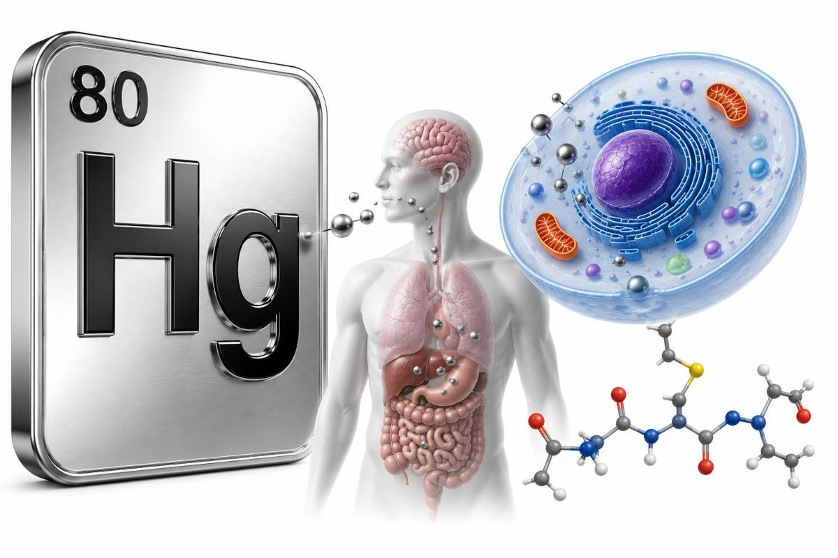
Mercury does not travel as a free ion in the body but instead primarily binds tightly to sulfhydryl groups (-SH) in proteins and enzymes, blocking their function and promoting oxidative stress.
Given the tendency of mercuric ions to form strong bonds with thiols, one can assume that all mercuric ions within biological systems are bound to thiol-containing molecules, such as glutathione (GSH), cysteine (Cys), homocysteine (Hcy), N-acetylcysteine (NAC), and albumin.
Mercury (specifically methylmercury and mercuric ions) acts as an ionic and molecular mimic in the human body by forming complexes with amino acids like cysteine. This mimicry enables mercury to hijack transport systems intended for essential nutrients.
Key pathologies of Mercury Mimicry- Amino Acid Transporter Hijacking:
Mercury gains entry into cells by mimicking natural amino acids like methionine and cystine.
After binding to sulfur-containing amino acids such as cysteine, mercury forms complexes that closely resemble essential nutrients recognized by cellular transport systems.
For example, the methylmercury–cysteine (MeHg–Cys) complex mimics the essential amino acid methionine.
This molecular mimicry allows it to cross the blood-brain barrier (BBB) through the L-type neutral amino acid transporter (LAT) system.
Mercury exploits cells through three primary mechanisms:- Thiol conjugation: binding to sulfur-containing molecules such as cysteine.
- Structural mimicry: forming complexes that resemble naturally occurring amino acids.
- Transporter hijacking: using amino acid transporters to gain entry into cells.
References:- Mercuric Conjugates of Cysteine Are Transported by the Amino Acid Transporter System b0,+: Implications of Molecular Mimicry (Bridges et. al., 2008)
"results implicate a mechanism of molecular mimicry, with Cys-S-Hg-S-Cys mimicking the amino acid cystine at the site of system b0,+."
- The methylmercury-L-cysteine conjugate is a substrate for the L-type large neutral amino acid transporter (Zhaobao Yin et al., 2008)
- Methylmercury transport across the placenta via neutral amino acid carrier (Y. Kajiwara, 1996)
- Developmental Toxicity: The methylmercury-cysteine complex passes through the placental barrier, leading to higher accumulation in fetal blood compared to maternal blood, increasing risks to the developing fetus.
References:- Mercury and Prenatal Growth: A Systematic Review (Dack et al., 2021)
"Mercury may be a threat to the developing foetus because both elemental and organic forms of mercury can cross the placenta during gestation, where it may accumulate in a far higher dose-to-weight ratio than is possible in an adult."
"Overall, studies mostly reported no strong evidence of an association between prenatal mercury exposure and birth weight"
"Almost all studies reported no strong evidence that prenatal mercury exposure is associated with birth length (10 of 14 studies), or head circumference (13 of 14 studies)."
- Methylmercury and Human Embryonic Development (M. Mandana, 2016)
"During the third week of gestation, the human nervous system begins to form in the embryo. During this gestational period, the embryo's nervous system is particularly susceptible to the influence of neurotoxins like methylmercury that can result in abnormalities."
"They found that the infants had higher concentrations of methylmercury in their blood than did their mothers, indicating that methylmercury easily transfered from pregnant women to their fetuses or embryos."
- Mercury toxicokinetics of the healthy human term placenta involve amino acid transporters and ABC transporters (Straka et. al., 2016)
"The findings can well explain why mercury is transported primarily towards the fetal side."
- Mercury and Prenatal Growth: A Systematic Review (Dack et al., 2021)
- Systemic Damage: Ionic mimicry contributes to the accumulation of mercury in the kidneys, liver, and blood, leading to tubular dysfunction, nephrotic syndrome, and cardiovascular issues.
References:- Mercury Exposure and Health Effects: What Do We Really Know? (Charkiewicz et al., 2025)
"Mercury and its forms, even in the smallest doses, cause numerous disorders to the body, including to the nervous system, the respiratory system, and the cardiovascular system. It can cause disorders such as various cancers; endothelial dysfunction; gastric and vascular disorders; liver, kidney, and brain damage; hormonal imbalances, miscarriages, and reproductive disorders; skin lesions; vision damage; and even death."
- Mercury Exposure and Health Effects: What Do We Really Know? (Charkiewicz et al., 2025)
- Lysosomal dysfunction: mercury interferes with interaction between the protein vesticle and the lysosome which supports the process of autophagy, resulting in impaired protein recycling. Failed tau protein recycling is implicated in Alzheimer's disease and failed alpha-synuclein protein recycling has been implicated in Parkinson's disease.
References:- Heavy metal-induced disruption of the autophagy-lysosomal pathway: implications for aging and neurodegenerative disorders (2025, Das et. al.)
"As individuals age, the consequences of neurodegeneration become more significant, raising the likelihood of developing disorders like Alzheimer's and Parkinson's disease. This review explores the intricate relationship between heavy metal exposure, dysfunction of the autophagy-lysosomal pathway, ..."
- Autophagy in Neurodegenerative Diseases and Metal Neurotoxicity (Ziyan Zhang et. al., 2016)
"PD is a late-onset neurodegenerative disease characterized by progressive and extensive loss of dopaminergic neurons in the substantia nigra pars compacta (SNpc) and the presence of aggregated a-synuclein-containing intracellular inclusions known as Lewy bodies. Lysosomal depletion and accumulation of autophagosomes are observed in the postmortem brain samples from PD patients, suggestive of defective autophagic clearance."
- Heavy metal-induced disruption of the autophagy-lysosomal pathway: implications for aging and neurodegenerative disorders (2025, Das et. al.)
- Inhibition of dopaminergic neurotransmission: Signaling from one motor neuron to the next requires the flow of neurotransmitters (eg. dopamine) via the synaptic clef. This is impaired by mercury.
References:- Methylmercury induces neurite degeneration in primary culture of mouse dopaminergic mesencephalic cells (Gotz et al., 2002)
"The morphometric analysis of DA neurons exposed to 1 microM MeHg demonstrated a striking decrease in the number of neurites, indicative of cytoskeletal alteration. In addition, DA neurons displayed cell shrinkage and a significant increase of nuclei with chromatin condensation. Based on these results it is concluded that MeHg is highly toxic to primary DA neurons."
- Methylmercury Impairs Canonical Dopamine Metabolism in Rat Undifferentiated Pheochromocytoma (PC12) Cells by Indirect Inhibition of Aldehyde Dehydrogenase ( Tiernan et al., 2015)
"MeHg does not directly impair aldehyde dehydrogenase (ALDH) enzymatic activity, however MeHg depletes cytosolic levels of the ALDH cofactor NAD+, which could contribute to impaired ALDH activity following exposure to MeHg."
"MeHg alters cellular and biochemical processes in a variety of cell types, including both astrocytes and neurons."
- Methylmercury induces neurite degeneration in primary culture of mouse dopaminergic mesencephalic cells (Gotz et al., 2002)
- Impairment Of Motor Control By Presence Of Mercury In The Substantia Nigra: The Substantia Nigra is the dark mid-brain region where the neurons are responsible for motor control.
This region is dark due to neuromelanin which is pigmented by the presence of sulfur.
Sulfur attracts mercury which is the same principal as a mercury chelator.
Mercury in turn interferes with operation of the motor neurons.
The following paper attributes this behavior to "higher vulnerability of the pigmented neurones" rather than the mechanism elucidated here.
References:- Substantia nigra neuromelanin: structure, synthesis, and molecular behaviour (Zecca, Tampellini et al., 2001)
- Impairment of Vision by Mercury: A study showed the toxic effects of mercury on the retinal nerve fiber layer thickness (RNFLT), macular thickness (MT), and choroidal thickness (CT).
Comparisons were made between those who experienced chronic exposure to mercury to a control group who was not.
The study referenced deficits in color vision in the blue-yellow range, visual field defects, absent or blunted corneal sensations, iris atrophy, pallor (becomes pale) of the optic disk and what seems like an endless list of optical maladies.
See study: Toxic Effects of Chronic Mercury Exposure on the Retinal Nerve Fiber Layer and Macular and Choroidal Thickness in Industrial Mercury Battery Workers (Ekinci et al., 2014)
This ionic and molecular interaction can lead to detrimental effects in many organ systems. These include, but are not limited to, the cardiovascular (endothelial dysfunction), gastrointestinal (nausea, vomiting, severe abdominal pain, and diarrhea), neurological (brain and nervous system), immune/autoimmune (for example skin rashes), hepatobiliary (liver) and renal (kidney), systems.
The effects of organ dysfunction can lead to misery and death.
More on mercury:
For more information on mercury, mercury detoxification and the pathology of mercury see the link between mercury and Parkinson's disease.
Other mercury topics on this website:
- Mercury measurement, chelation and the pathology of mercury
- Project to develop a new Mercury Chelator
- Project to investigate new techniques to measure the body burden of mercury
- Dental amalgam removal and the IAOMT SMART protocol
References: